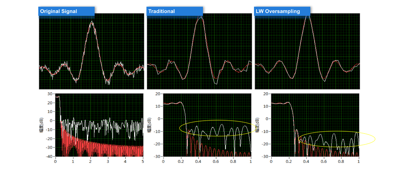
Patent Signal Oversampling
Landwind Medical won patent on Signal
Oversampling. This technology can effectively avoid the two-dimensional image
distortion brought by linear interpolation of the Digital Signal
Controller,remove the noise outside of the blood flow, improve the sensitivity
of theblood flow detected, and Increase the grayscale image resolution
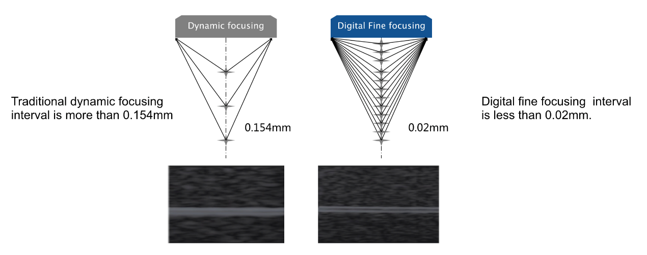
LW Digital Fine Foucsing
The interval of traditional dynamic
focusing is more than 0.154mm while digital fine focusing interval is
less than 0.02mm.
Each sample point can be precisely
focused.
Effectively improve the superficial
image resolution
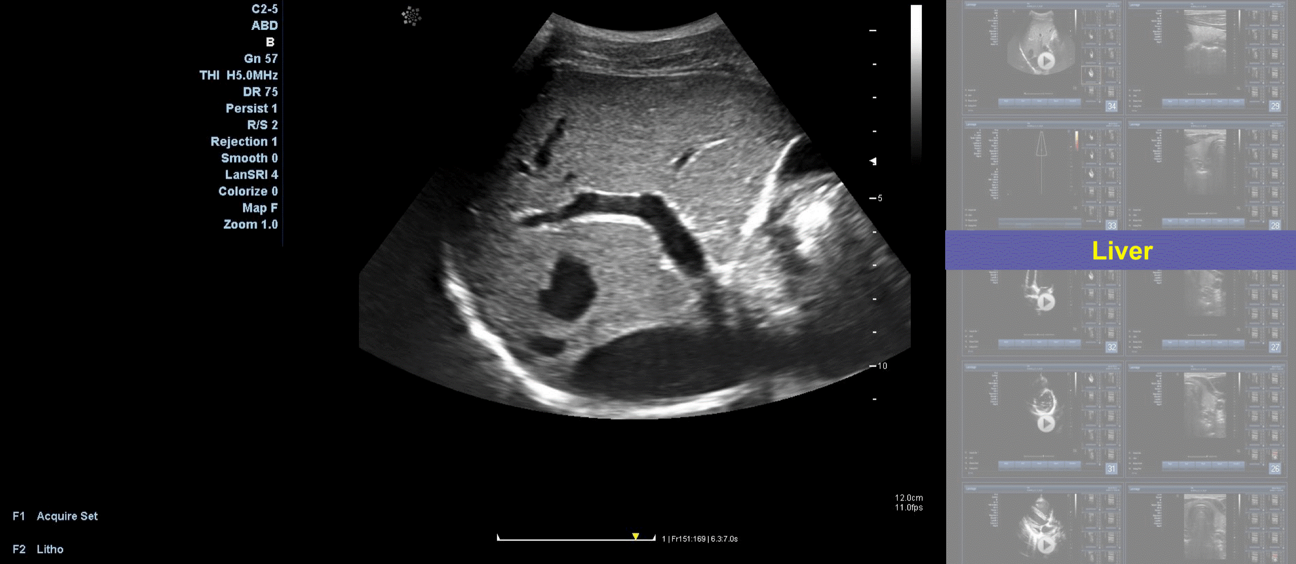
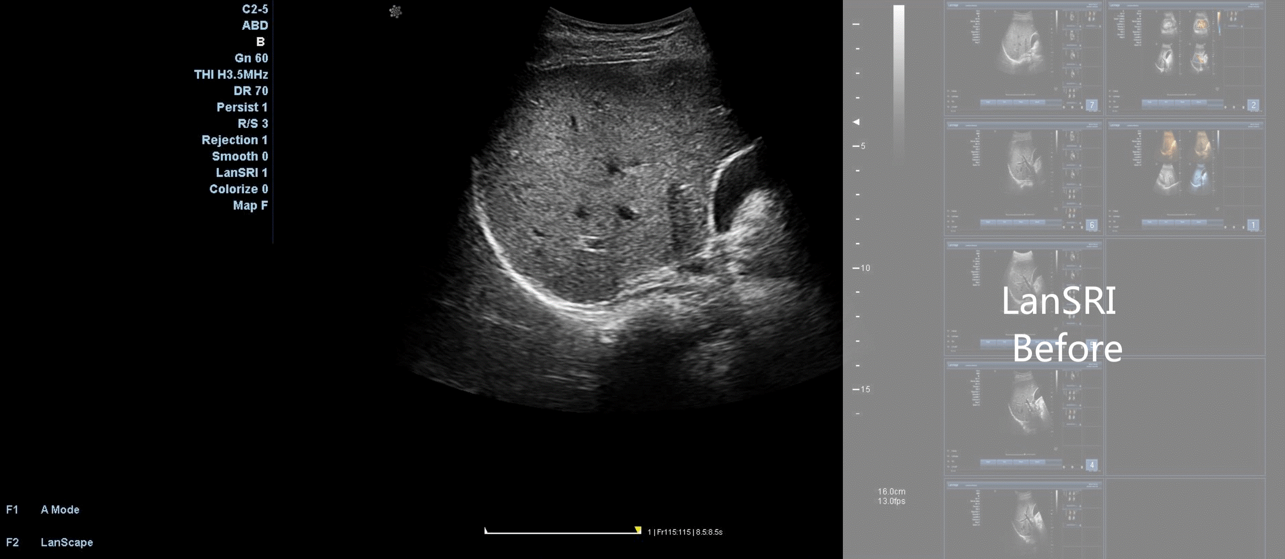
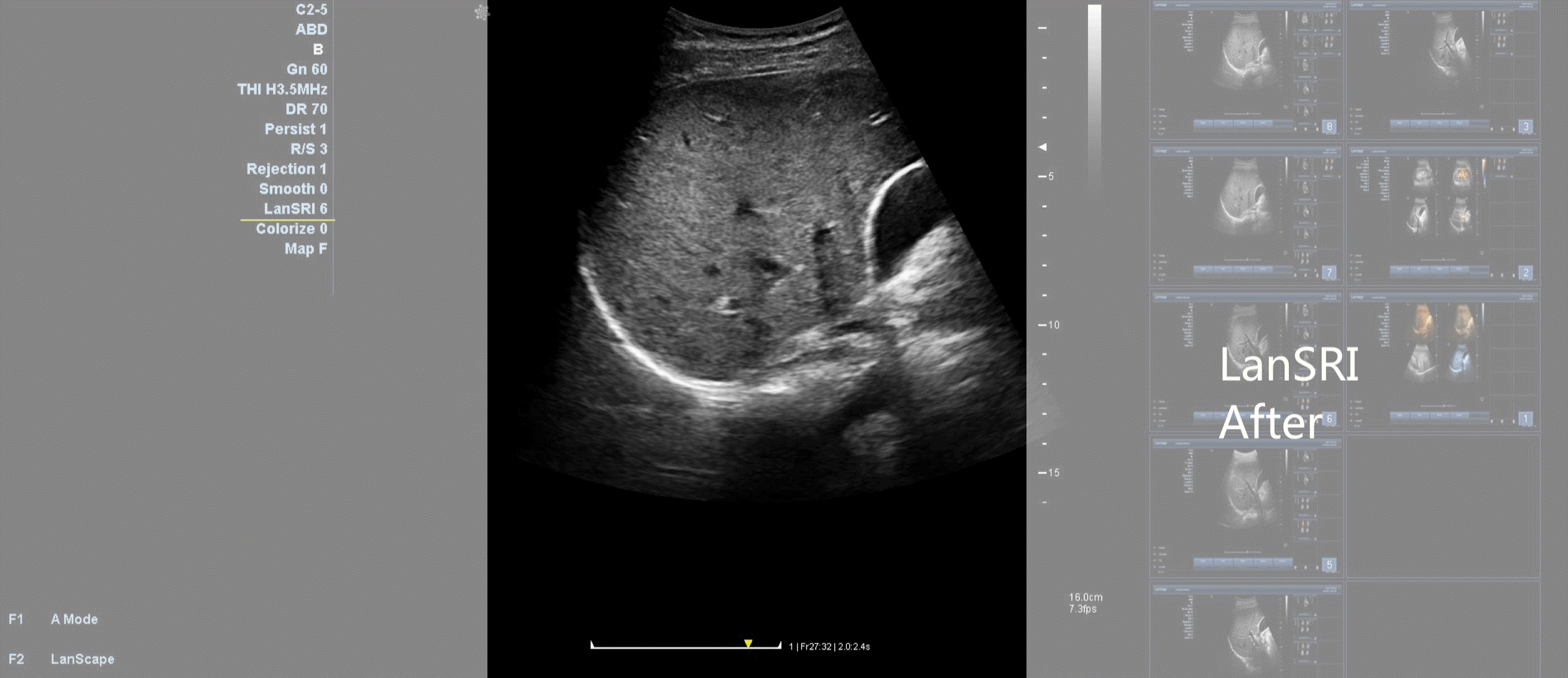
LanSRI: Speckle Reduction Imaging
By LanSRI we mean decompose the image to a
group of smaller ones in pyramid structure, analysis the texture information to
remove speckle noise in different scales and then reconstruct the image after
the noise suppression. LanSRI can help to enhancesmooth along preferred
edge direction, reduces variation within speckle region, and maintain local
mean gray scale level.
LanSRI can be turned on/off and 4 levels
adjustable on:
ϙ live
images
ϙ frozen
images
ϙ images
recalled from the archive
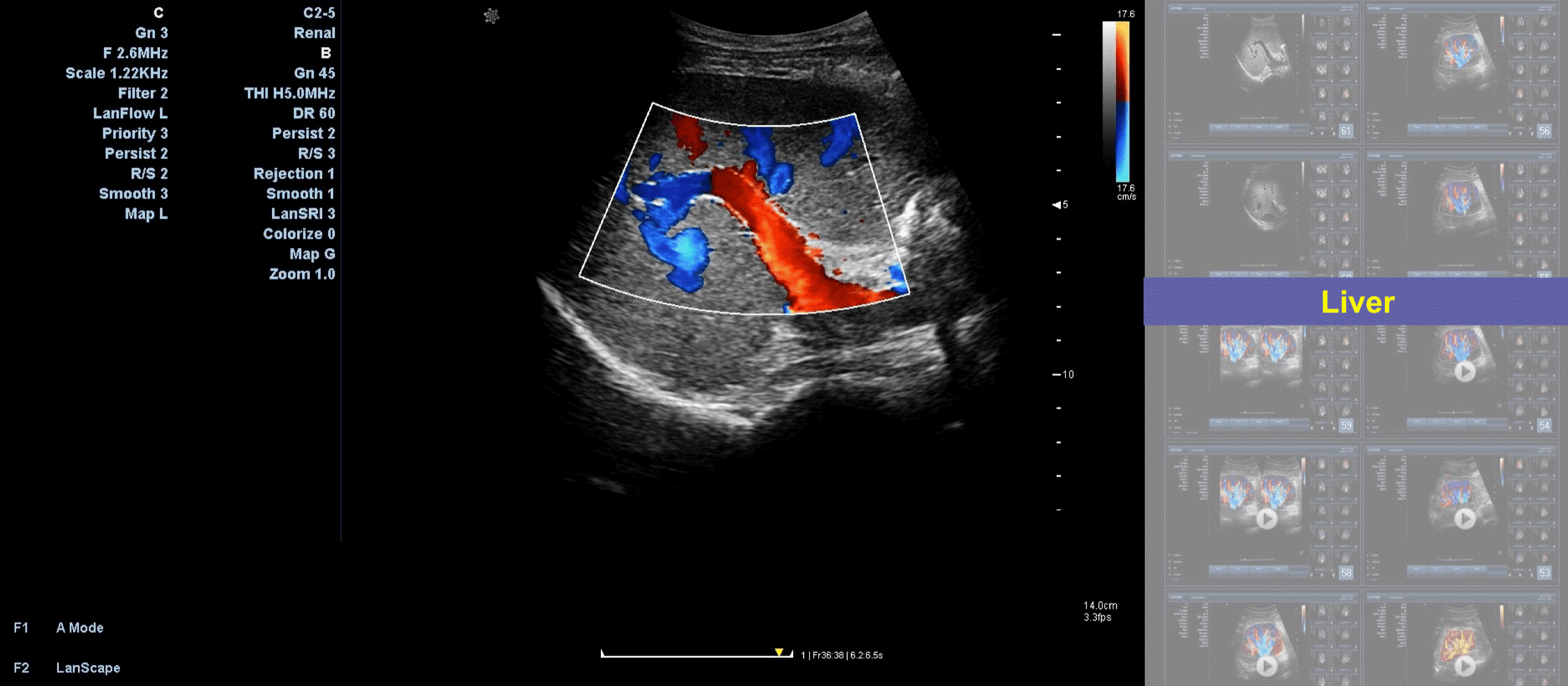
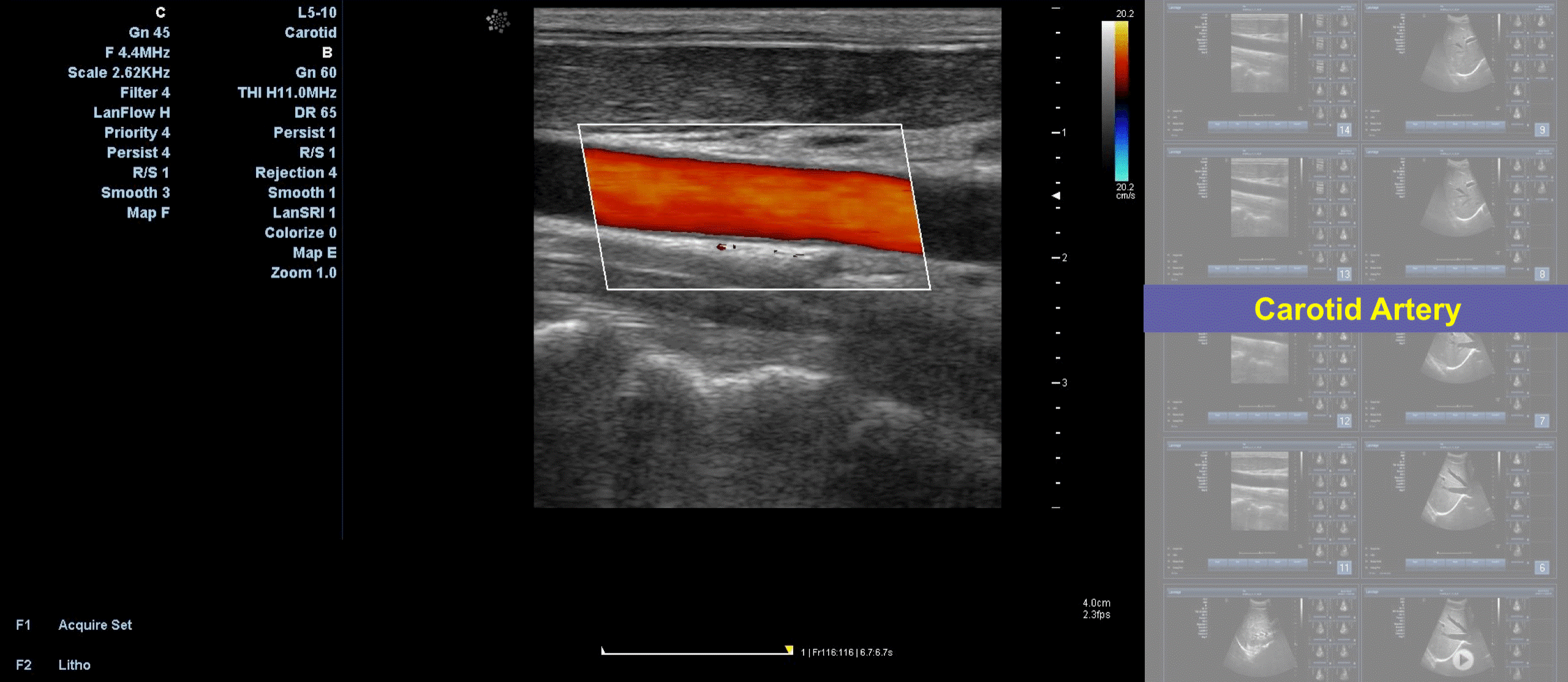
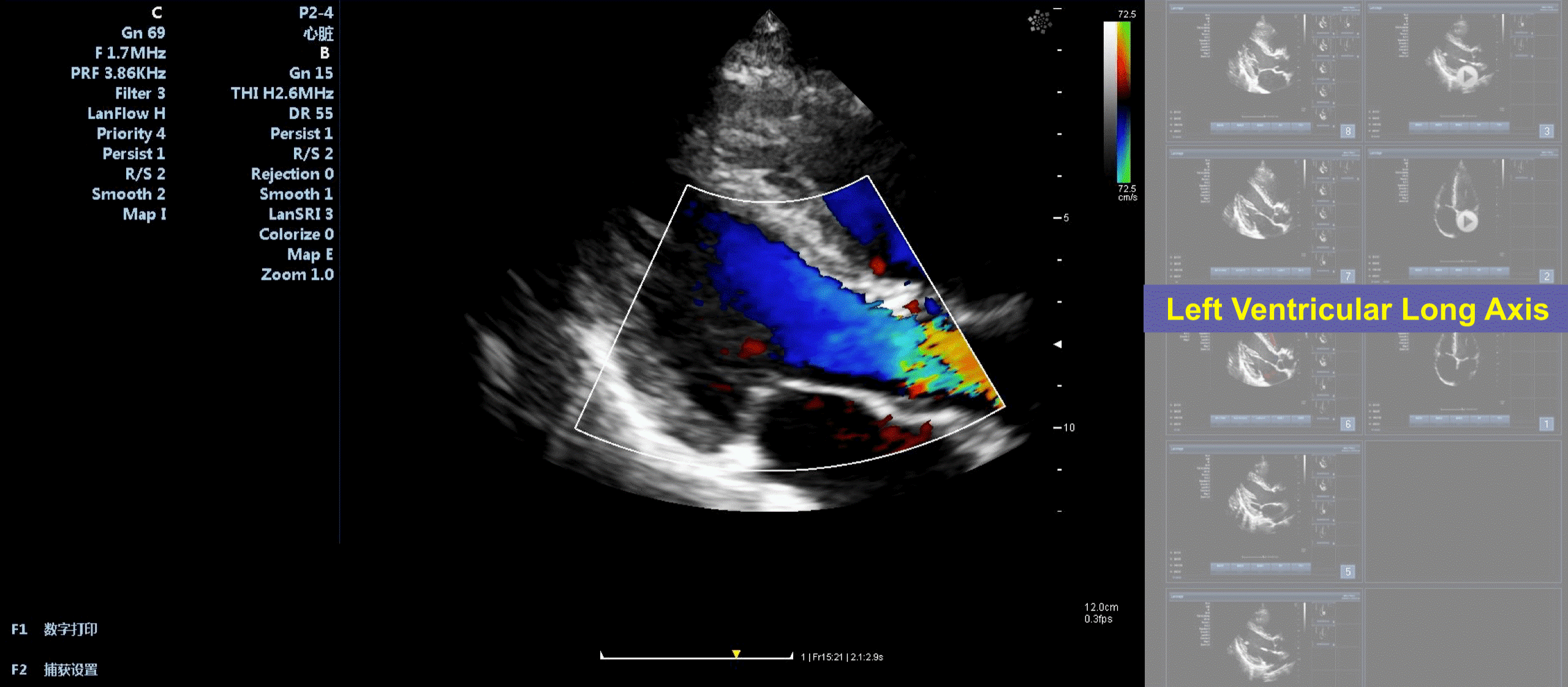
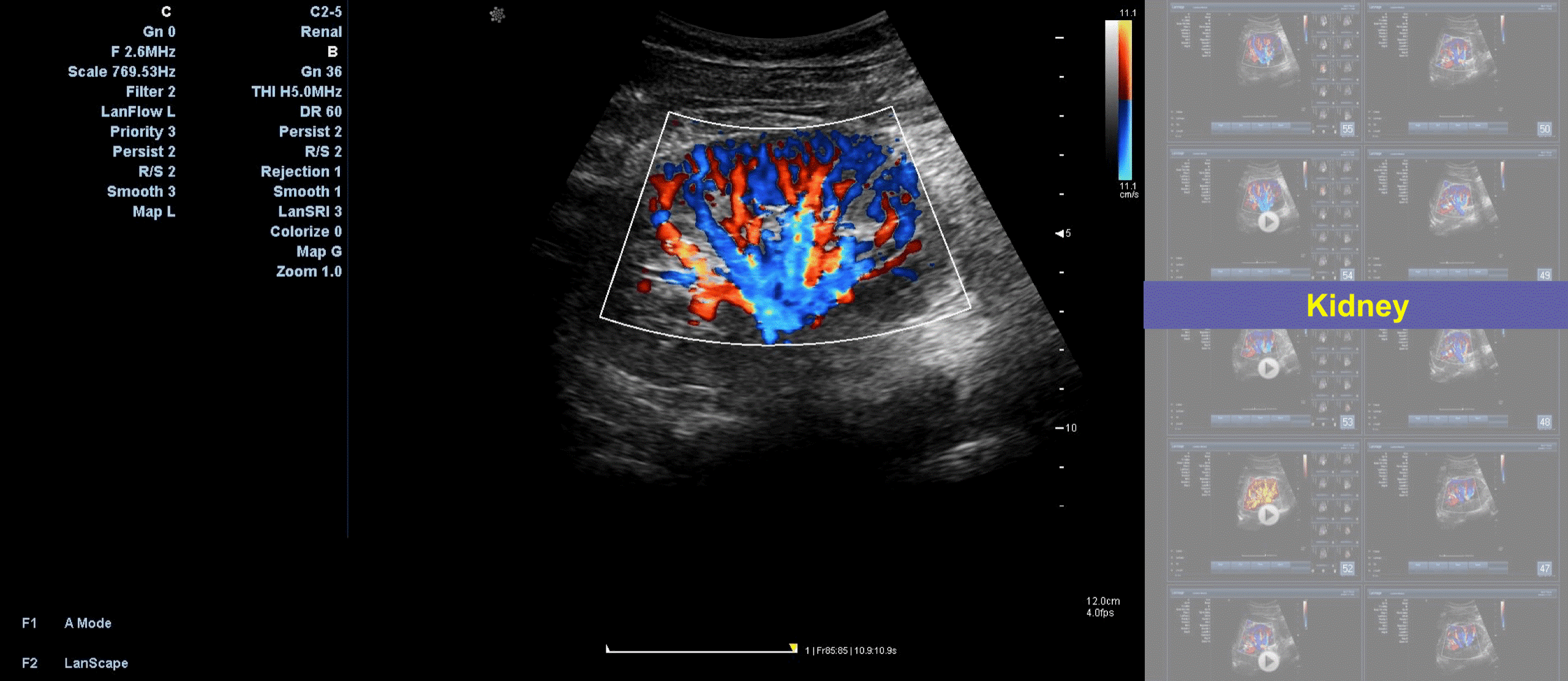
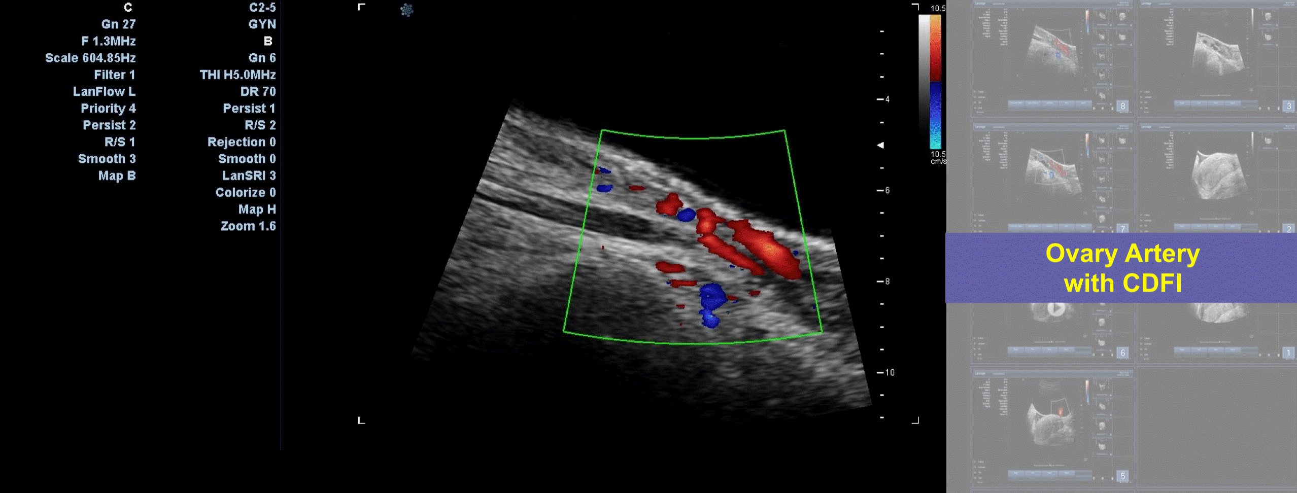
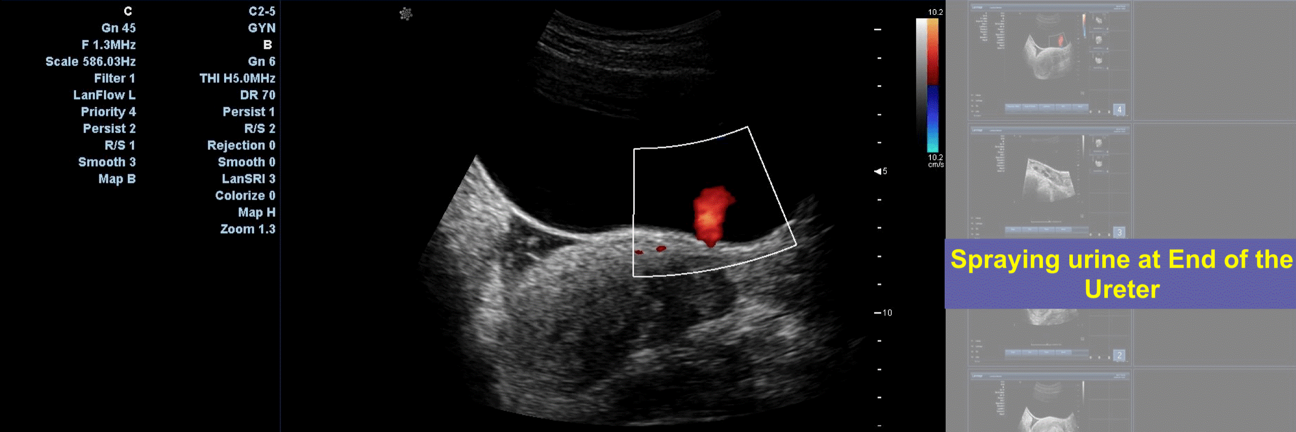
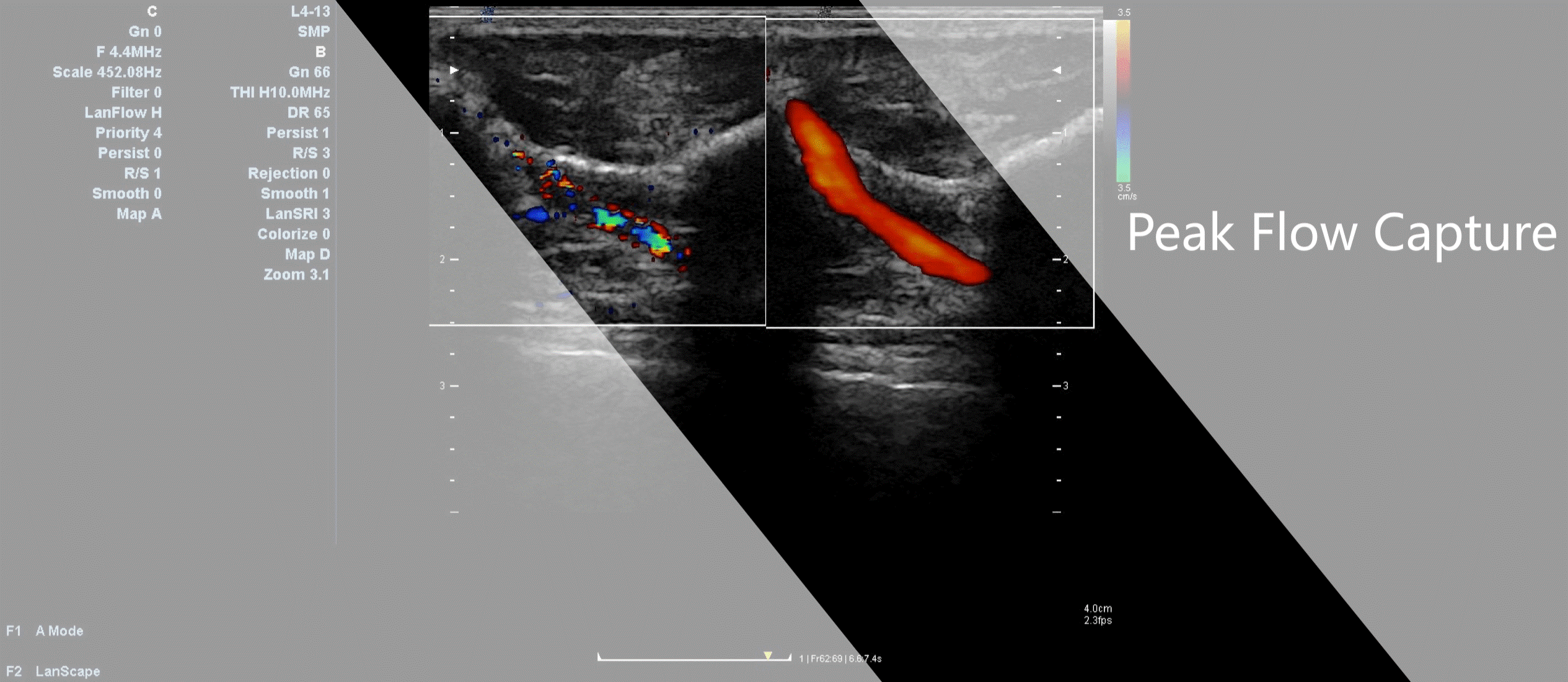
Color Flow Peak Velocity Capture
Capture the peak flow signals within a
certain time duration, in order to improve the low speed blood flow and low
perfusion organ such as tumor blood supply.
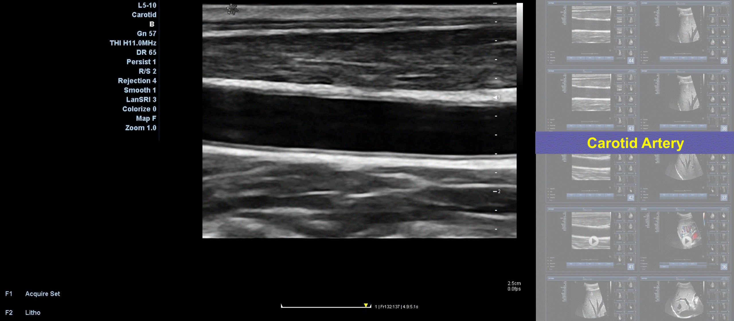
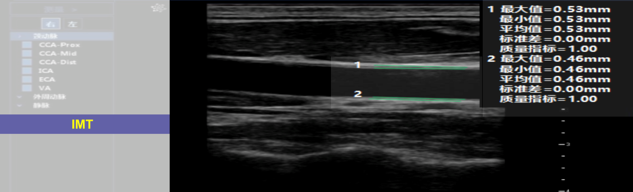
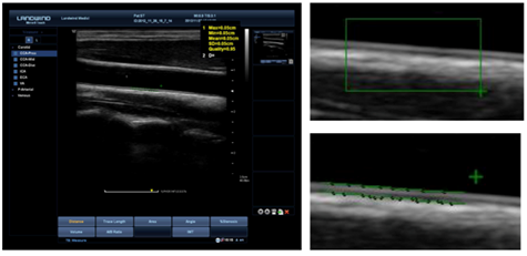
Auto IMT
Automated measurement of the intima-media
thickness of the carotid artery wall, for evaluating an asymptomatic patient’s
risk of developing cardiovascular disease.
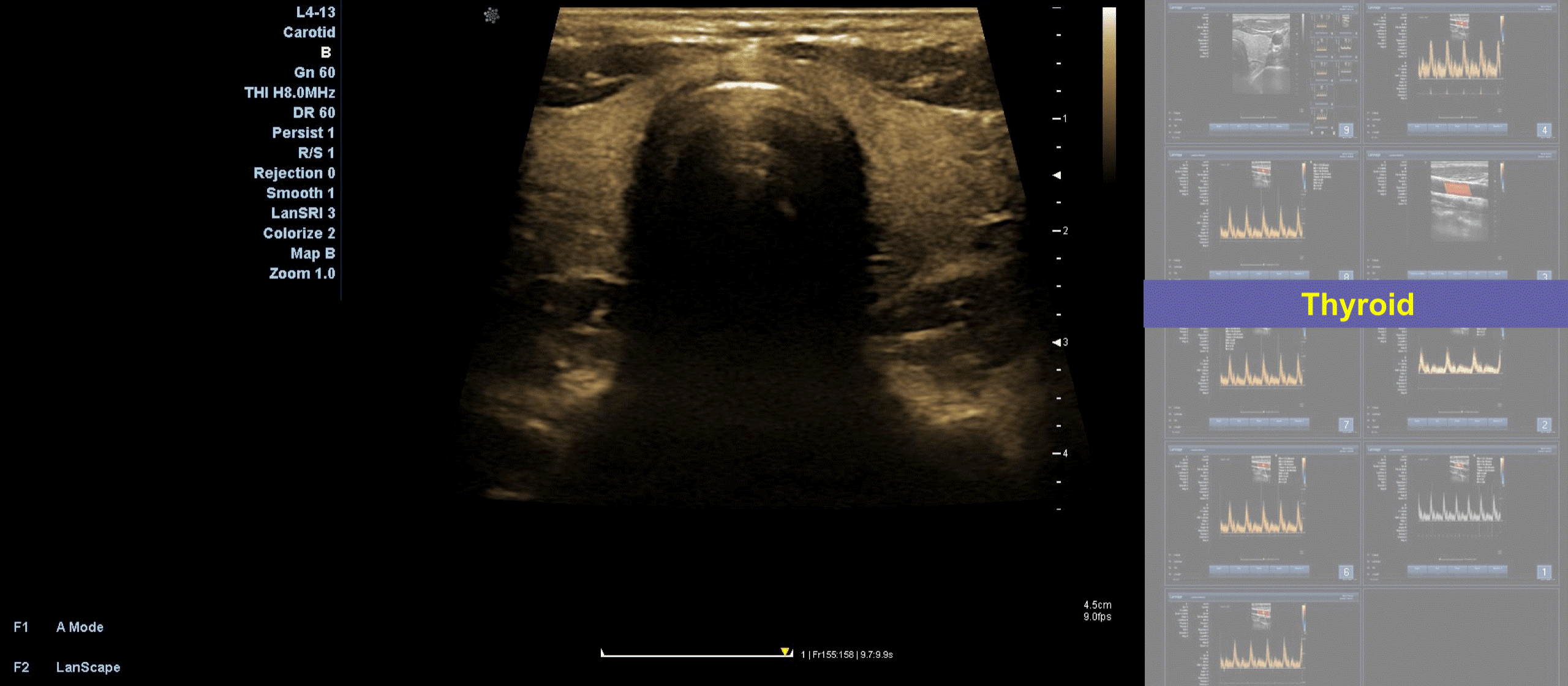
Tissue Specific Imaging
Sound Speed varies in different
tissues.Tissue Specific Imaging enables doctors to adjust the speed of sound
according to patient condition so as to
minimize beam distortion and improve imaging
results and diagnostic outcome. It has 4 adjustable levels: General, MSK, Fluid
and Fat.
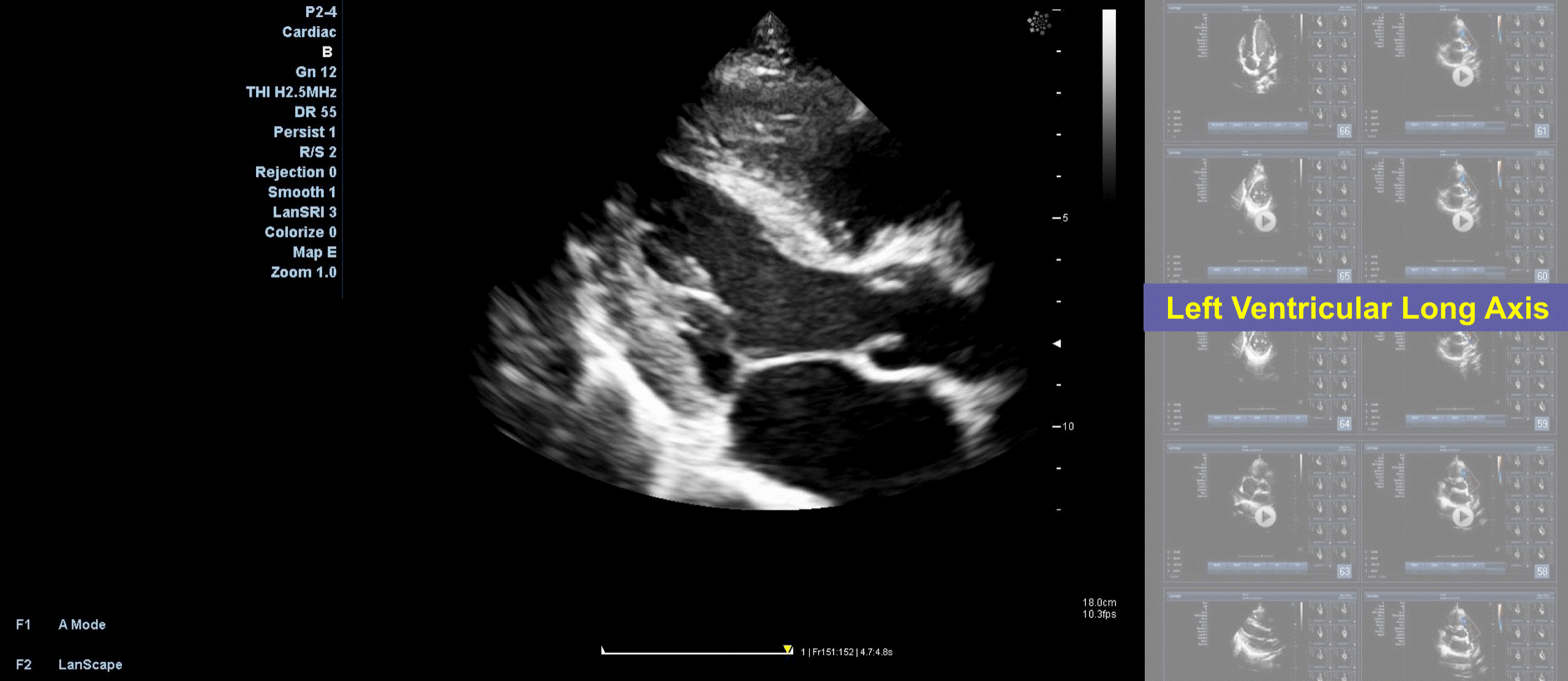
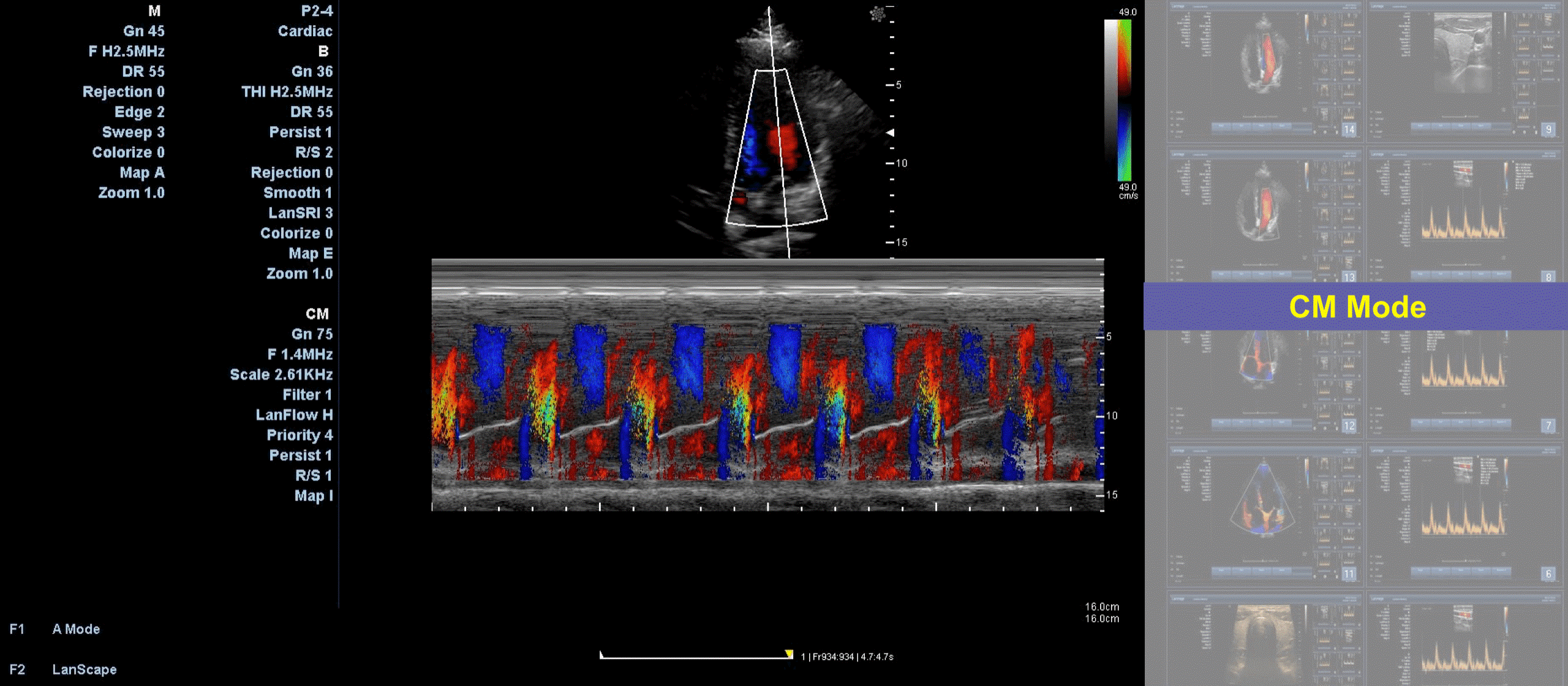
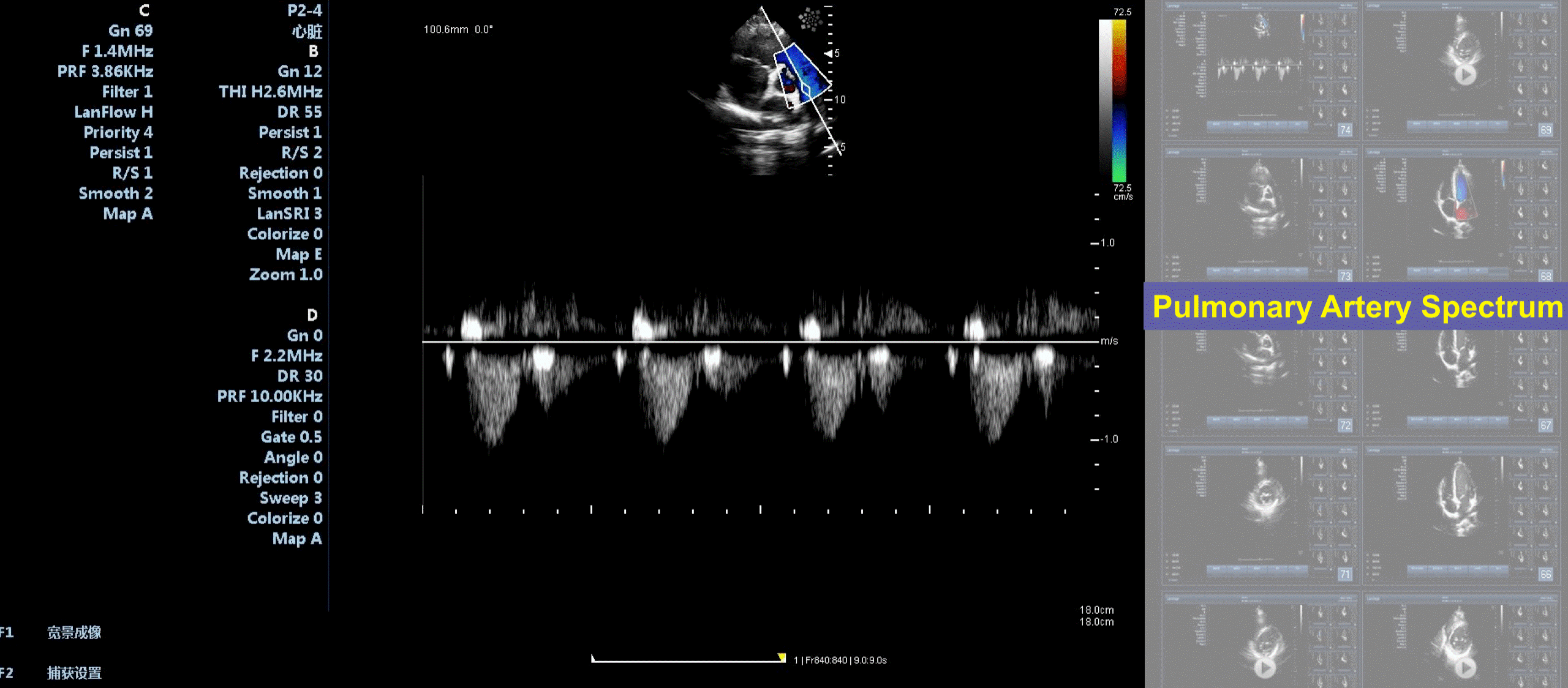
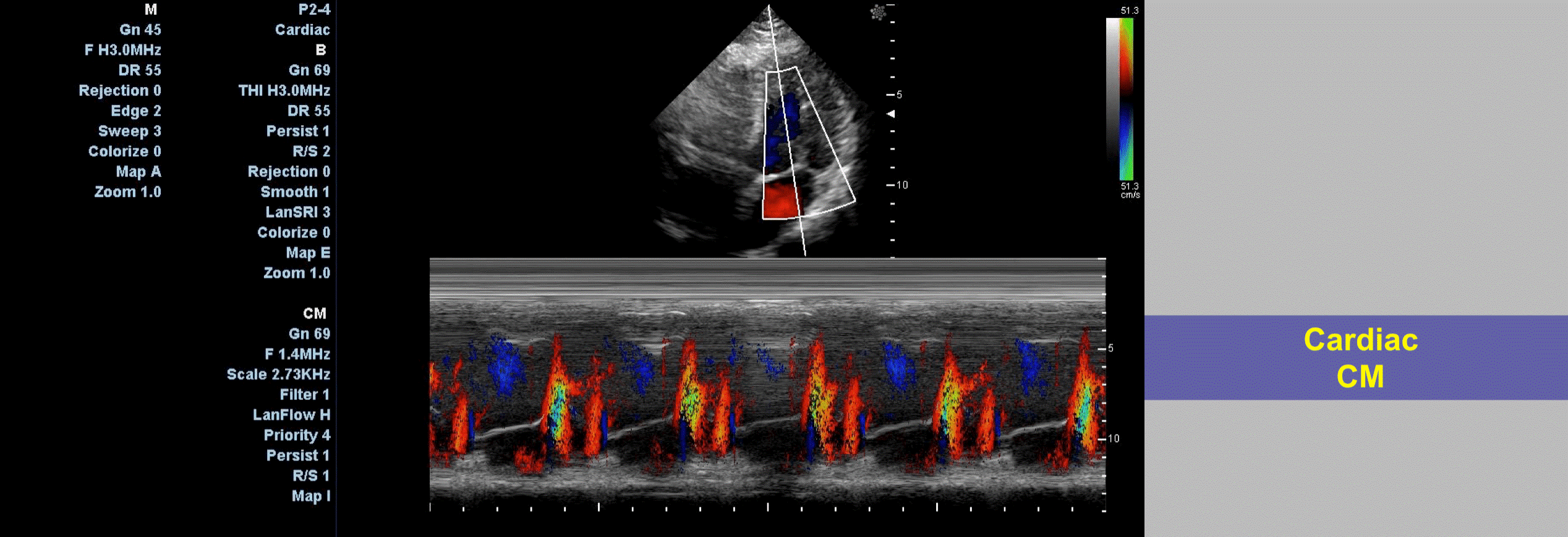
Color M Mode
Color m-mode imaging is a technique which
uses pulsed Doppler interrogation along a single line of interrogation, similar
to M-mode echocardiography.
In color m-mode Doppler, the Doppler
velocity shift is recorded and then color encoded and superimposed on the
M-mode image. This process results
in high temporal resolution data on the
direction and timing of flow events. Color M-mode (CMM) echocardiography provides a spatiotemporal map
of
the velocities of the blood flow along the
scan line from the mitral annulus to the LV apex.
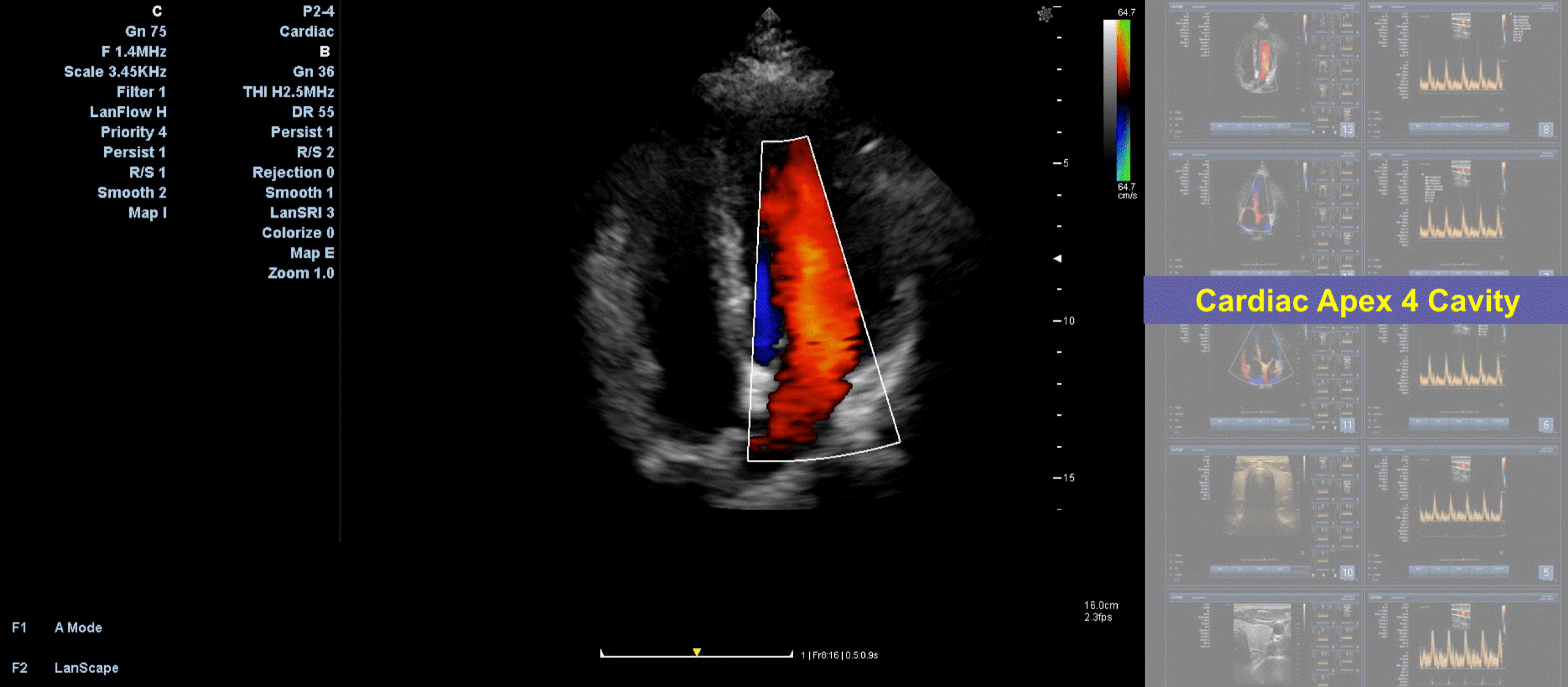
Tissue Velocity Mapping
Tissue Velocity Mapping is also called
Color-coded Tissue Doppler Imaging. It can create two dimensional color images
that facilitate visual assessment
of ventricular wall more significantly.
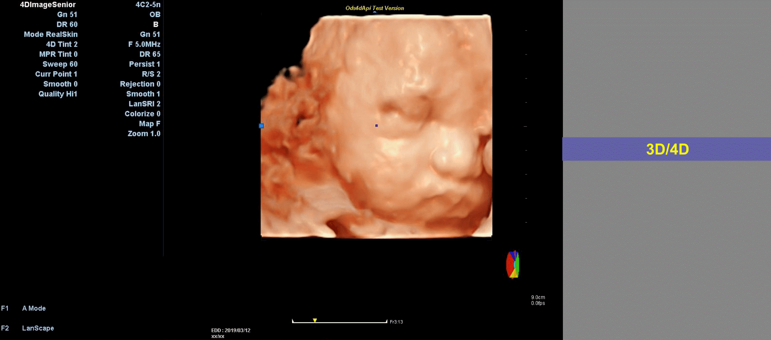
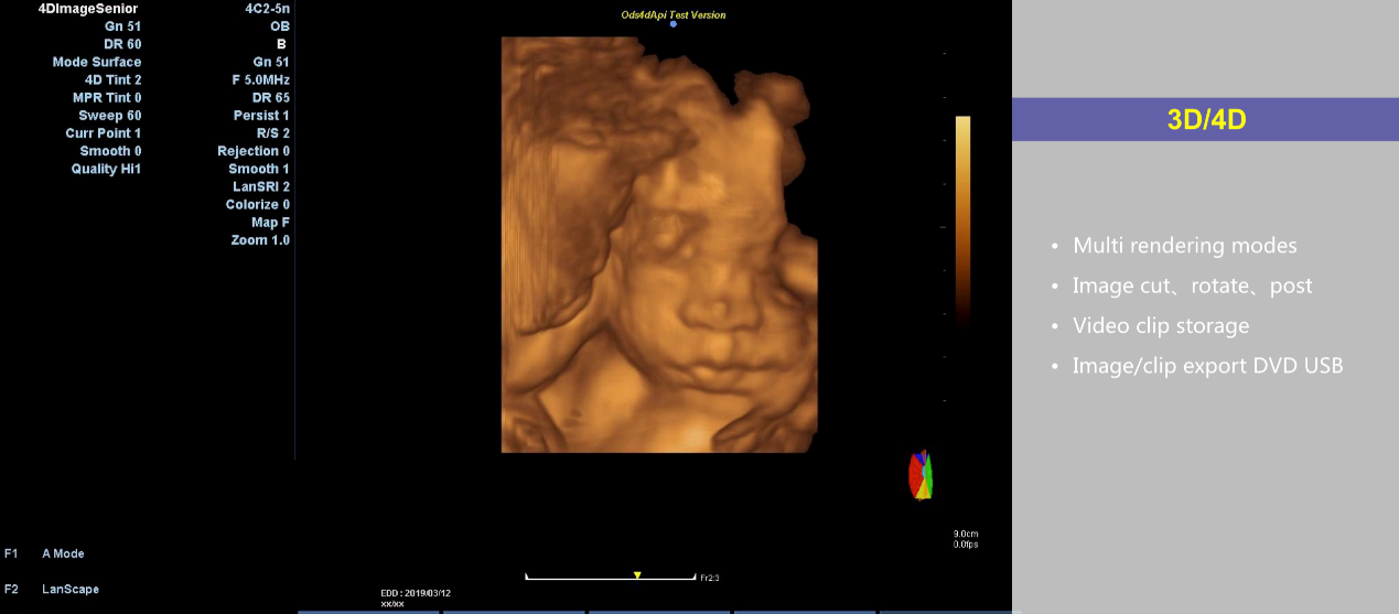
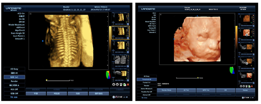
4D: LanSkin
Landwind Medical’s 4D package are capable of
advanced 4D capabilities like LanSkin andTomographic Ultrasound Imaging.
ϙ LanSkin:
Real Skin Imaging with light source, depth view
ϙ Tomographic
Ultrasound Imaging: function like CT Scanning
Elastography
Ultrasound Elastography is a medical imaging modality
that maps the elastic properties of soft tissue.Whether the tissue is hard or soft will give
diagnostic
information about the presence or status of
disease, providing a color map of tissue elasticity that is superimposed on the
real-time grey scale ultrasound image.
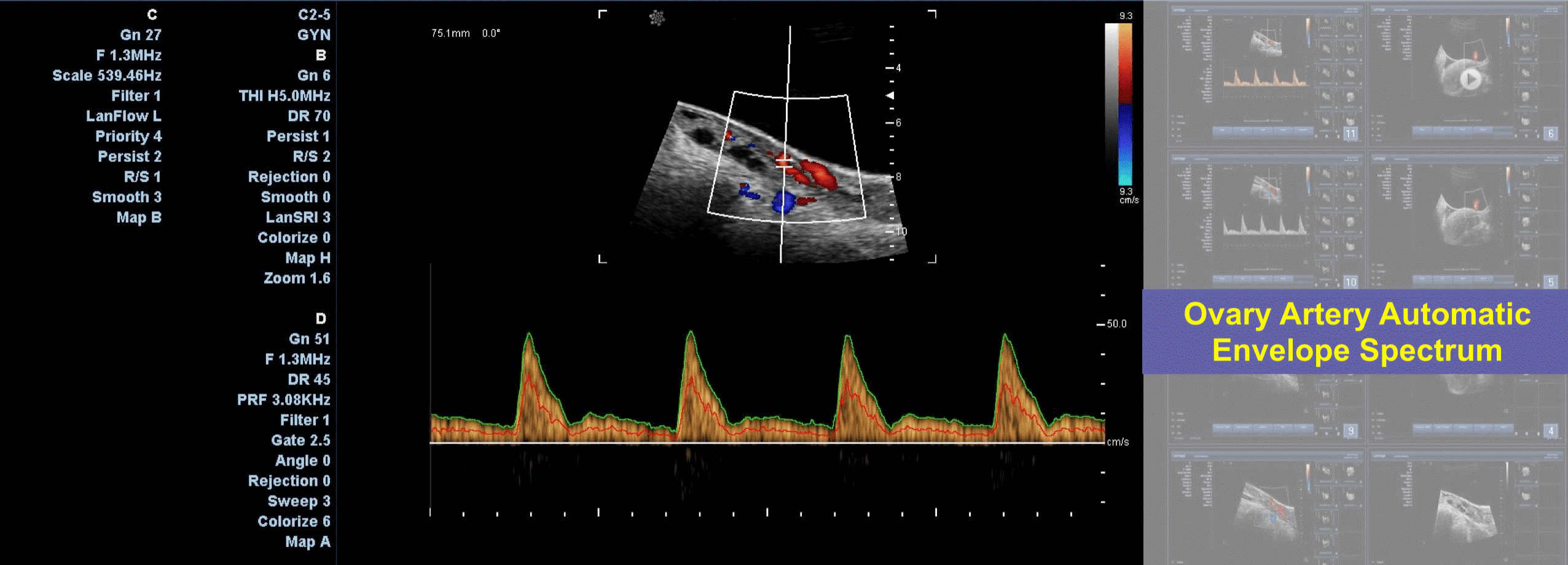
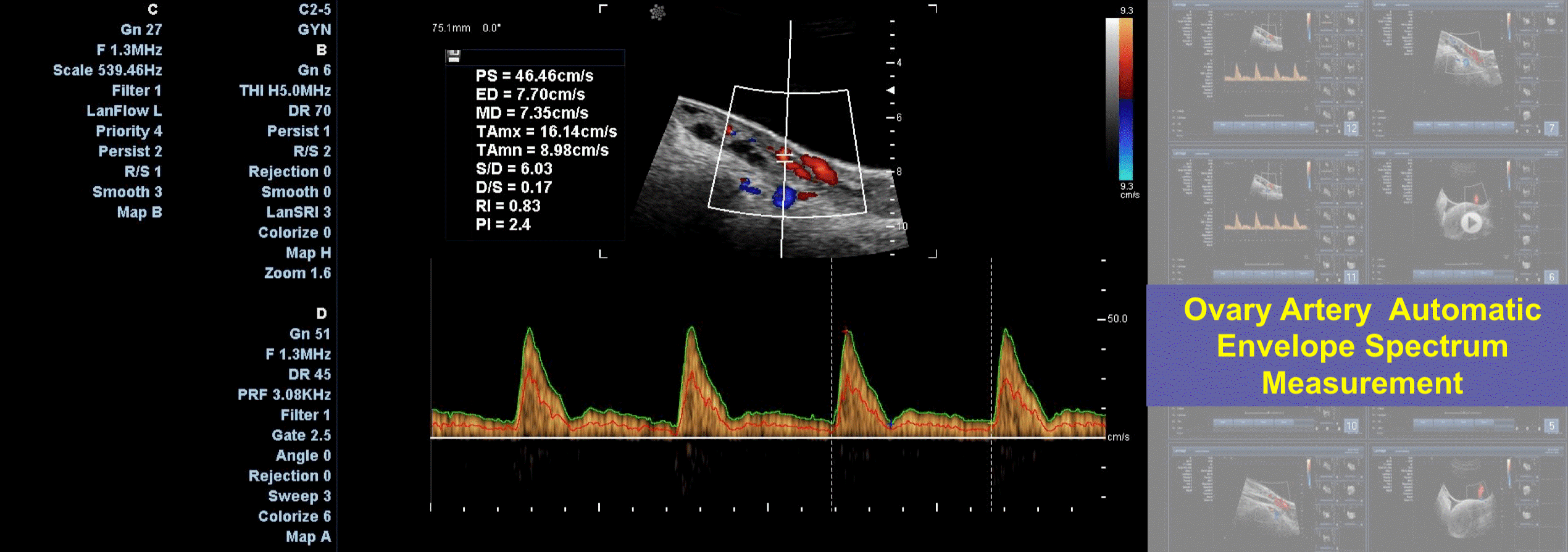
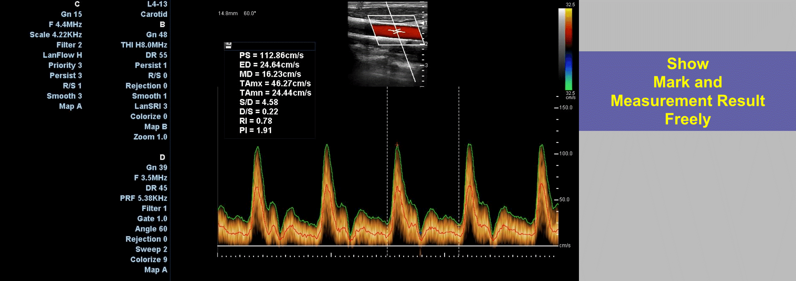
Auto Trace and Auto Calculation
By Auto trace and auto calculation, doctors
can get necessary parameters automatically.
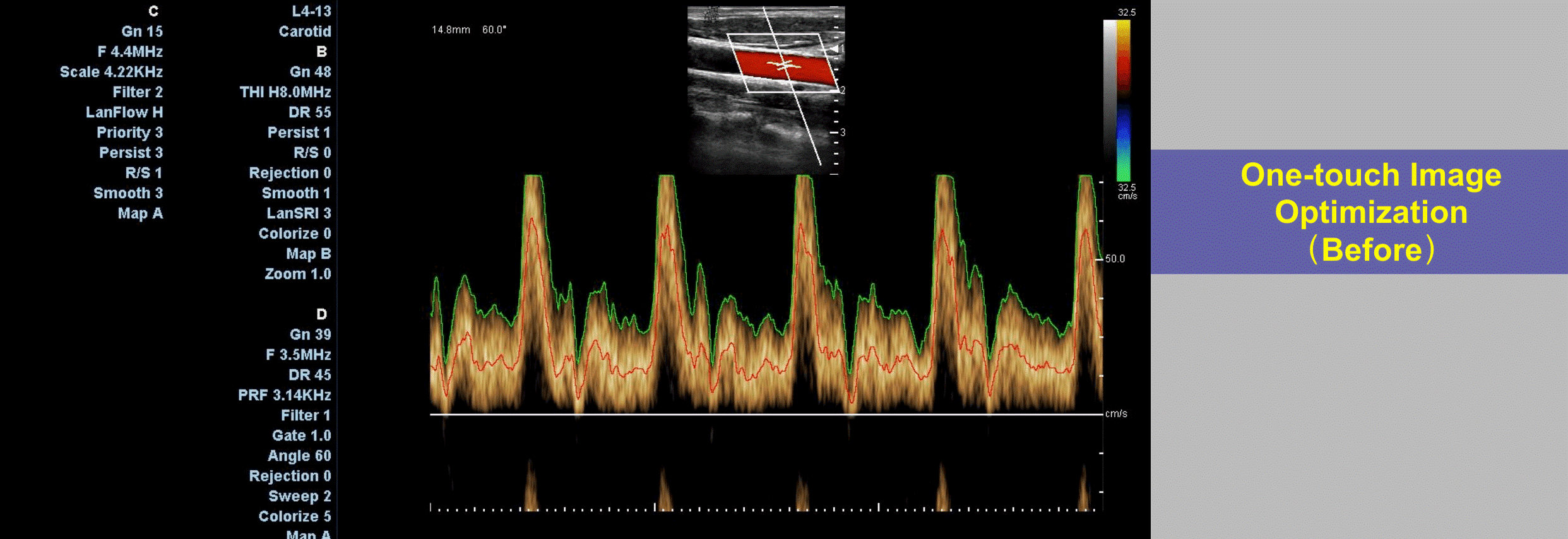
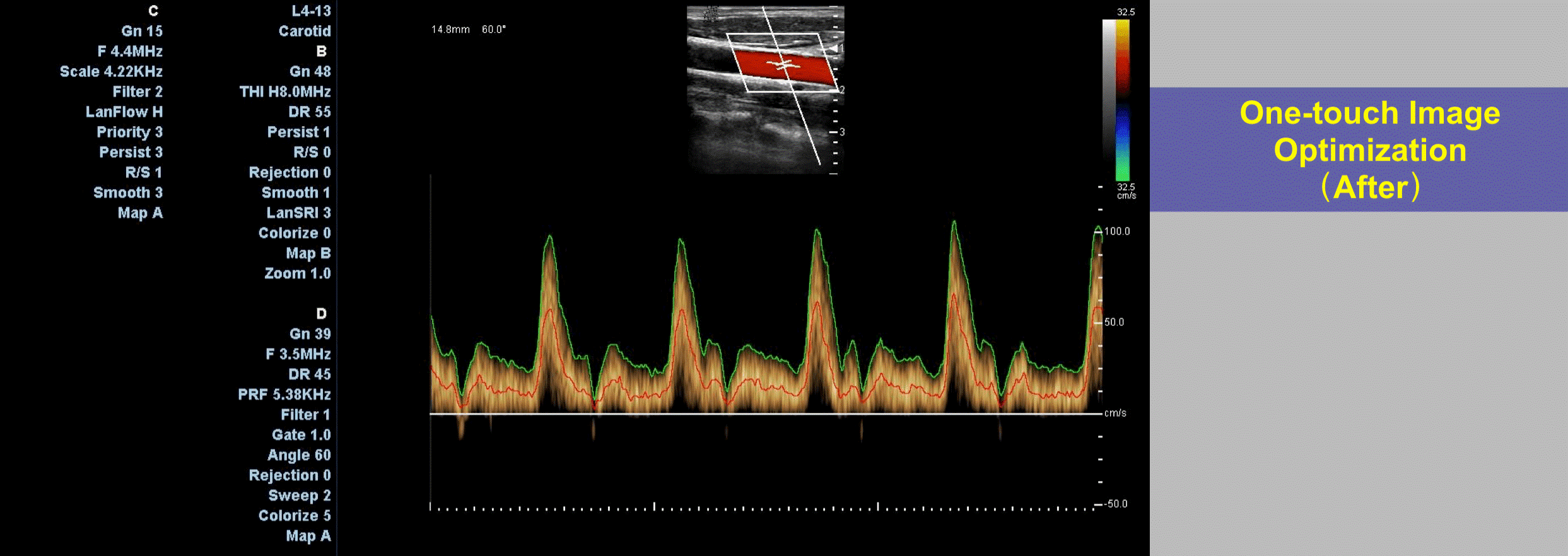
One-touch Workflow:
ϙ one-touch
Image storage, export, print
ϙ One-touch
export DVD USB
ϙ One-touch
Image Optimization
ϙ one-touch
spectrum Optimization