-

Landwind Medical is a health care company headquartered in Shenzhen, China with its own modern Research & Development Centers and Manufacturing facilities to ensure high quality products and services to our customers.
-

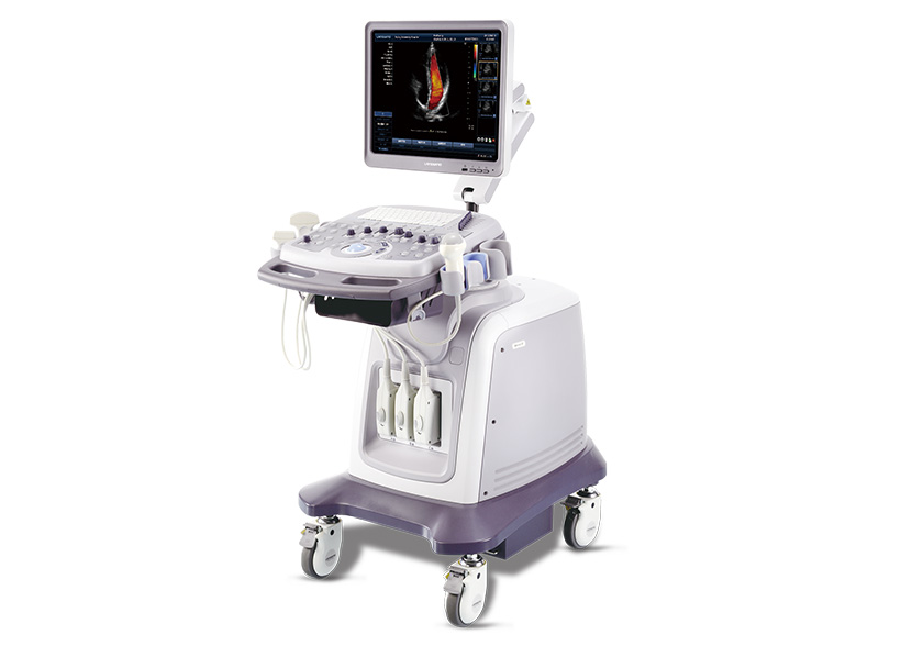
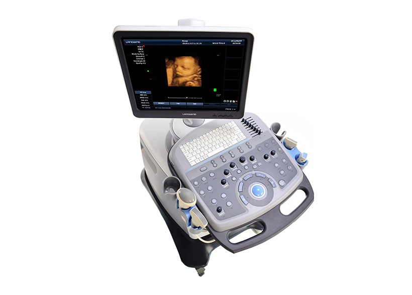
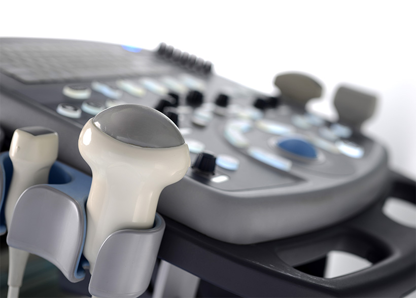
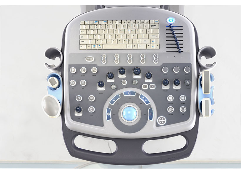
5 product lines meet your various needs. They are:Ultrasound machines and probes, IVD machines and reagents, Radiology machines and solutions, Anesthesia machines, Ventilator, and Hemodialysis products and solutions.
-

More than 20 years' experience. Two separate teams to support both domestic and international market. We are 365 days and 24 hours available for all partners. English is our working language.
-

What do you want from your job? Do you just want a paycheck or more than that?
At Landwind Medical, you will discover more about your potential, push your limits, and grow yourselves with Landwind Medical.
-

Shenzhen Landwind Biomedical Technology Co., Ltd
Add: Block B15, Life Science Park, 140 Jinye Avenue, Dapeng New District, Shenzhen 518000, Guangdong, CHINA.
E-mail: sales@landwindmedical.com
Tel: +86-755-8393 5015